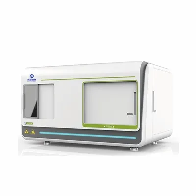
CellScan - Live Cell Imaging System
CellScan
The CellScan Live Cell Imaging System can be placed in the incubator together with the living cells to form an in-box experimental center. During the entire culture process, the cell growth status can be obtained in real time through remote monitoring, and the cell number and confluence can be calculated using the AI intelligent analysis system, which
greatly saves time and economic costs in traditional cell culture methods, and helps reseachers to dynamically understand the health status, morphology and function of cell models more efficiently.
Description
Easy Operation
Compact structure, space saving
CellScan integrates the microscopic imaging and illumination optical systems inside the device, making the instrument compact and saving space in the incubator.

Simple process management
From observation project establishment to data export, a process management model is adopted. The operation interface is simple and easy to use, truly realizing simpler cell observation.

Anytime anywhere, remote viewing
CellScan builds a local area network through a router, and uploads data to the cloud server. Users can log in
through the cloud platform account to browse remotely and obtain images and quantitative data anytime and anywhere.

AI-driven, real-time quantification
Multiple built-in cell models allow for easy identification of the shape of specific cell types in changing cell states
and automatic calculation of cell counts and confluence in real-time. Machine
learning based methods allow users to manually annotate cellular and non-cellular regions to build specific analysis
models for fusion analysis.

Application

Cell confluence degree is directly related to cell proliferation and activity. Starting from the cell fusion curve, the effects of different culture factors, serum concentrations or drug toxicity and other variable factors can be evaluated.


Customize imaging parameters
- Multipoint imaging: Customize the sites to monitor based on your area of interest, or use simple, fast pre-set templates for imaging.
- Focus mode: The autofocus mode uses artificial intelligence to identify the best field of view, and can still obtain the clearest image in long-term monitoring experiments. The manual focus mode can customize the focus height of each observation point.
- Brightness adjustment: Auto/manual exposure.
- Monitoring time interval: The shortest time interval can be set to 5 minutes.
For downstream experiments such as cell passaging and transfection that require accurate judgment of cell confluence, users can set their own confluence thresholds and choose email or SMS notifications.

Compatible with various culture vessels
CellScan is suitable for culture dishes, culture bottles, multi-well plates and multi-layer cell factories, and is compatible with more than 30 different brands and more than 600 culture consumables.

Autofocus, maintain optimal view
The system performs imaging with an AI-driven smart recognition algorithm that grabs the optimal focus height point and maintains the clearest field of view over days, weeks, and even months of long-duration experiments.

Using non-invasive analysis methods, high-order AI algorithms automatically identify cells and clusters, quantify the cellular regions on the container surface, count the number of cells and the degree of fusion in real time, and generate time-lapse videos, cell count scatter plots, and confluence curves at the end of the project.

Comparative data analysis
Data comparison of monitoring sites on the same or different devices facilitates growth curve analysis between duplicate wells and various experimental treatment factors.

Hot Tags: CellScan - Live Cell Imaging System, China CellScan - Live Cell Imaging System manufacturers, suppliers, endoscopic imaging system, water maze test, Digital Pathology Slide Scanner, radial arm maze test, Auditory development, Laser speckle contrast imaging system
Send Inquiry
You Might Also Like