Key Factors Determining the Quality of Digital Pathology Slide Scanners
Leave a message
Key Factors Determining the Quality of Digital Pathology Slide Scanners
The quality of a digital pathology slide scanner can be encapsulated into three primary areas:
Resolution and Lighting Technology:
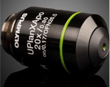
The G-Cell digital pathology slide scanners are equipped with cutting-edge technologies such as high-speed stroboscopic light sources, apochromatic flat-field microscopy, and Köhler illumination. These advanced features collaborate to deliver ultra-high-resolution optical imaging, providing crisp and detailed pathological images that serve as a reliable basis for accurate diagnoses.


Motor Control Technology:
The precision of movement in a digital slide scanner is paramount. G-Cell scanners incorporate piezoelectric ceramic stack nano-stages for minute adjustments, a lead screw motor for the z-axis, and linear motors for the XY-axis. This combination ensures ultra-high-precision motion control, guaranteeing stability and accuracy throughout the scanning process, ultimately enhancing image quality.

Algorithms and Image Processing:
The integration of sophisticated algorithms and software is crucial for efficient image processing. G-Cell scanners boast intelligent recognition algorithms and fast autofocus technology, enabling high-speed processing. They also feature field curvature digital correction and wide-range focal plane fitting, ensuring that regardless of specimen flatness, clear images are captured in every field of view. Real-time image stitching enables the seamless creation of complete specimen images, while digital resolution enhancement and realistic color restoration techniques contribute to the production of high-quality images that are both visually appealing and diagnostically valuable.


In conclusion, the G-Cell digital pathology slide scanners excel in resolution and lighting technology, motor control technology, and algorithms for image processing, making them a formidable tool for supporting accurate and reliable pathological diagnoses.


